Research
We build microfluidic platforms and engineered soft-matter objects to answer quantitative questions in biology, from single molecules to whole organisms.
Our work sits at the interface of microfluidics, soft matter, biophysics and immunology, with a strong emphasis on droplet-based systems. We generate, manipulate and functionalise droplets, interfaces, particles and cells to create controlled microscale environments, and combine microfabrication, interface chemistry, optical microscopy and quantitative image analysis to turn them into robust tools for biological questions.

Axis 1
Functional droplets and particles as biological tools
Emulsion droplets and microparticles with controlled size, softness and surface chemistry, used as artificial targets to measure how immune cells sense, engulf and process what they meet.
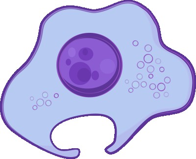
Biomimetic droplets to decode phagocytosis
How do the size, deformability and surface chemistry of a target control the way macrophages engulf and degrade it?
Biological targets vary in many properties at once. We replace them by lipid emulsion droplets whose interface we functionalise with defined ligands (antibodies, mannose, biotin) and fluorescent reporters, so that one parameter can be changed at a time. With this toolkit we showed that target size alone tunes phagocytic uptake, built FRET-based particles that report receptor clustering at the interface, and ratiometric particles that follow phagosomal acidification quantitatively, cell by cell.
Key result Target size alone controls phagocytic uptake — Biophysical Journal (2019)
Funding ANR Imperis with P. Pierobon (Institut Cochin, coordinator) and J.-F. Rupprecht (Centuri, Marseille)
Past funding ANR Phagochemiforce with F. Niedergang (Institut Cochin, coordinator), J.-M. Mallet and B. Dumat (CPCV, ENS)
Emerging direction
Lipid digestion at the droplet interface
Where and how fast are lipids digested, inside phagosomes or by gut microbes, and what limits this process?
Emulsion droplets are natural substrates for lipases. We are extending our droplet and imaging tools to measure lipolysis in space and time: inside the phagosomes of macrophages, and by microbial communities involved in fat malabsorption (with T. Le Roy and F. Carrière). Two MSCA postdoctoral projects are open on this theme — see open positions.
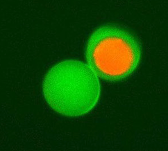
Degradable particles from vegetable oils
Can bio-sourced oils replace petroleum-based polymers for degradable, drug-loaded microparticles of controlled size?
We shape acrylated epoxidised soybean oil into monodisperse particles and capsules by microfluidic (double) emulsification and photo-crosslinking, and characterise how they degrade and release their cargo. The fabrication route is protected by a patent (WO2023237670).
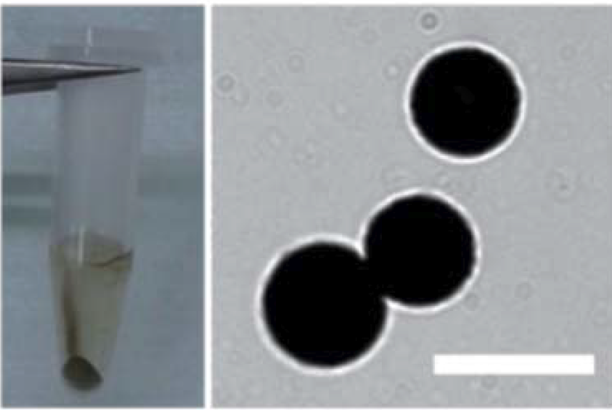
Coated droplets as optical and flow tracers
Can a liquid droplet carry a solid, functional shell, and serve as a tracer for flows?
Using emulsion droplets as templates, we showed that polydopamine coatings become fluorescent under UV, made metal-coated liquid particles for micro-velocimetry, and recently validated soybean-oil emulsions with red-blood-cell-sized droplets as a blood-mimicking fluid for both ultrasound Doppler and optical velocimetry.
Axis 2
Microfluidic platforms for quantitative cell biology
Trap arrays, droplet–cell pairing and reconstituted membrane systems that turn single-cell and single-vesicle observations into statistics.
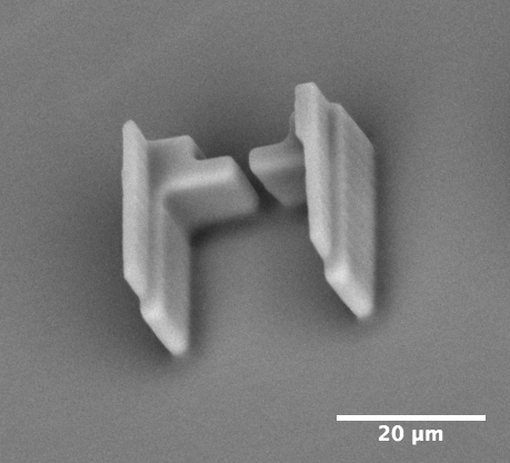
Trapping arrays for single-object statistics
How can we capture hundreds of cells, droplets or particles at defined positions, image them, and release them on demand?
We design hydrodynamic trap arrays and optimise their efficiency from the flow physics: oblique streams and lateral dispersion raise capture efficiency and homogeneity in dense arrays. Our latest device adds a pneumatically actuated membrane to release the trapped objects after analysis, opening the way to selection.
Key result Fully automated trapping, analysis and release — Micro and Nano Engineering (2025)
Immune cell polarity, one synapse at a time
How fast and how reproducibly do lymphocytes polarise when they meet an antigen-presenting surface?
By pairing single B cells with antigen-coated lipid droplets in microfluidic traps, we follow the formation of the immune synapse in many cells in parallel and extract quantitative polarisation dynamics (with P. Pierobon, Institut Cochin). The same approach revealed how microtubules restrict actin polymerisation to the synapse.
Key result Droplet–cell pairing platform — Cell Reports Methods (2022)
Reconstituted membranes under control
How do actin networks reshape lipid membranes, measured on whole populations of vesicles rather than a few?
With C. Campillo (Université d’Évry), we trap giant vesicles in a microfluidic device and sequentially expose them to actin machinery. This showed that branched actin networks stabilise lipid microdomains.
Axis 3
Microfluidics for plant and environmental biology
From the spatio-temporal study of moss development and microalgae phenotyping to the dynamics of hydrocarbon-degrading bacterial biofilms at oil–water interfaces.
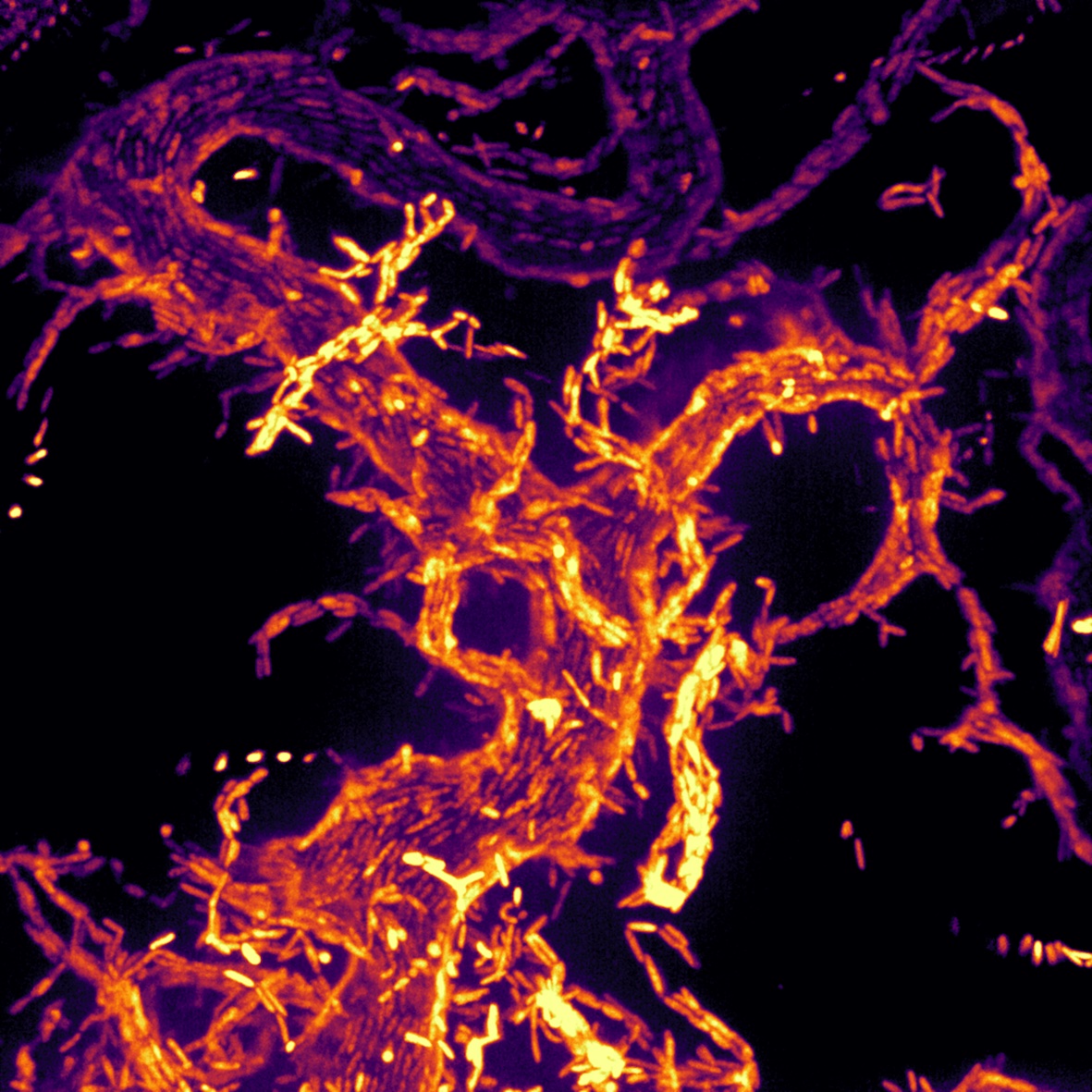
Bacteria that reshape oil to eat it
How do oil-degrading bacteria access hydrocarbons that are insoluble in water?
Alcanivorax borkumensis forms biofilms on oil droplets. With Andrew Utada (University of Tsukuba) and J.-F. Rupprecht (CPT, Marseille), we showed that these biofilms deform the oil–water interface into tubes, increasing the contact area and accelerating oil degradation — a mechanism relevant to the bioremediation of oil spills.
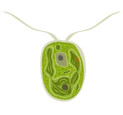
Selecting single microalgae
Can we phenotype individual microalgae and keep the interesting ones?
We develop automated microfluidic setups to immobilise single microalgae, measure optical readouts such as fluorescence, and release them for downstream culture.
Funding ANR Phosphalgue (coordinator) with L. Jullien (CPCV, ENS), A. Boulouis and S. Eberhard (IBPC), F. Guyot (MNHN), G. Charvin (IGBMC)
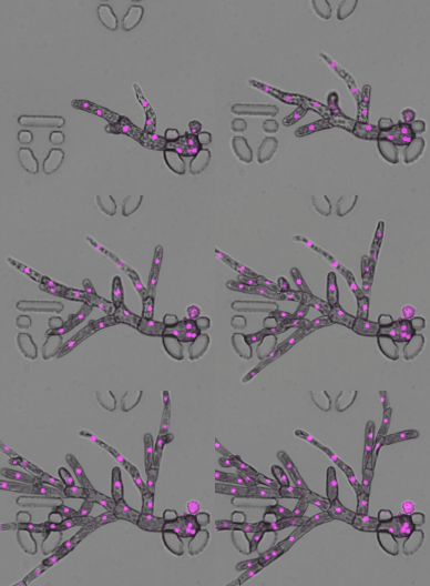
Plant development on chip
How does a single plant cell regenerate a whole organism, followed from seconds to weeks?
With J.-C. Palauqui (IJPB, INRAE Versailles) we built a microfluidic and microscopy toolbox to follow the moss Physcomitrium patens from single protoplasts to whole plants. We also contribute to quantitative pipelines for protoplast culture with S. Verger (UPSC, Umeå).
Translation: Quintessence Biotech
 Part of our work is translated through Quintessence Biotech, a start-up co-founded from the laboratory’s patent portfolio, which develops microfluidic and physico-chemical solutions for cell and gene therapies. This keeps a direct feedback loop between fundamental research, technology development and biomedical applications. More on LinkedIn.